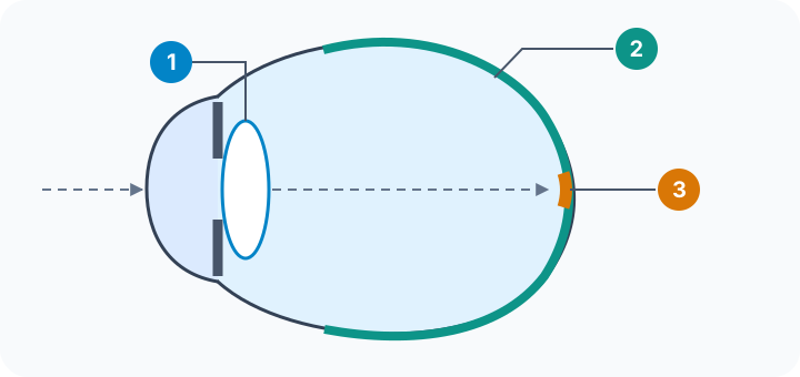
Location diagram only, not to scale, disease severity or a home vision test.
- 1 · Lens
Focuses incoming light near the front of the eye.
- 2 · Retina
Light-sensitive tissue along the back of the eye.
- 3 · Macula
A small retinal region important for central fine detail.
Seeing a cup is not the same task as reading its label
The lens near the front focuses light; the retina at the back turns light into signals. Within the retina, the small macular region is especially important for the fine detail in words and faces. AMD therefore differs from cataract, which concerns a cloudy lens. These conditions can coexist.
Imagine someone who can still locate a cup on a table but struggles to read the small print on a packet beside it. This illustrates how central detail can be impaired while some surrounding vision remains. It is not a symptom checklist or a test for AMD. Serious central loss can greatly restrict everyday life even without losing all vision.
Type and stage answer different questions
Dry and wet describe different disease changes. Dry AMD includes deposits called drusen and may involve thinning or loss of retinal support cells and light-sensitive cells. Wet, or neovascular, AMD involves abnormal fragile blood vessels that can leak fluid or blood and damage the macula. ‘Wet’ does not mean watery eyes; ‘dry’ does not mean harmless.
Early, intermediate and late describe the stage. Early AMD may have no noticeable visual loss; late disease can be dry or wet. Wet AMD is a late form and often affects vision faster, but dry AMD does not inevitably become wet. People and eyes can have different courses. Neither smoking history nor how well someone reads today identifies a stage.
What the smoking link can—and cannot—tell you
Age and inherited susceptibility also matter. Smoking is one changeable risk factor among them, not an explanation for every case or a way to assign blame. A study reporting fewer new AMD diagnoses is asking a different question from a study of progression in eyes that already have AMD. Neither outcome alone proves that quitting restores lost central vision.
An appropriately qualified eye-care professional can distinguish lens and retinal problems and explain the diagnosed type and stage. Useful questions include ‘Which part of the eye is affected?’ and ‘Does this name describe the type, the stage, or both?’ Qualified local cessation services have a separate role. The eye-care discussion concerns the tissue and diagnosed changes; cessation support concerns the process of stopping smoking. One does not establish the result of the other.
What to keep in mind
Sources
The central claims on this page were checked against the sources below.
- US National Eye Institute: AMD: central retinal anatomy, stages, smoking risk and qualified eye care
Sources checked: 2026-10-10
- 日本眼科学会 / 日本眼循環学会: 黄斑と水晶体の位置、新生血管型・萎縮型、喫煙と眼科専門職の役割
Sources checked: 2026-10-10
General eye-health education, not personal diagnosis, screening eligibility, home testing, symptom triage or treatment advice.