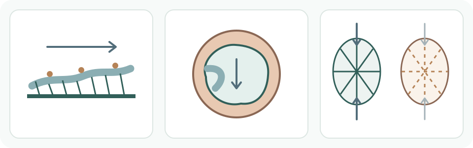
Surface transport, airway space and tissue recoil are different tasks. In the right-hand pair, solid support lines and inward arrows contrast with broken support lines and a lighter recoil arrow. This is a qualitative comparison, not a damage scale, personal stage or inevitable progression.
- Surface transport
Mucus and cilia move trapped material toward the throat.
- Airway space
Inflammation and mucus can reduce the passage for air.
- Tissue recoil
Loss of elastic recoil can make exhalation harder.
Airway clearance is a transport system, not a detox counter
Mucus traps some inhaled particles and cilia beat in a coordinated direction to move that mucus upward. Cough can assist this process, while particles that reach deeper regions involve other defenses such as macrophages. These mechanisms are related, not interchangeable.
The system does not create a visible daily score for how much smoke has left the lungs. A change in mucus or cough frequency cannot be converted into a toxin total, a percentage of damage or proof that the lungs are clean.
Smoke can affect cilia, mucus and airway lining at the same time
Authoritative reviews describe reduced ciliary function and impaired mucociliary clearance with cigarette-smoke exposure. Smoke can also change mucus production and the airway lining, so slowed transport is not simply a story about individual hairs stopping.
Mechanistic, laboratory and population evidence answer different questions. A laboratory observation can support biological plausibility, but it does not provide a personal dose meter or predict which symptom one person will notice.
Two ways air can become harder to breathe out
Mucus-and-cilia clearance moves material along the airway surface; ventilation moves air through the airway. They interact but are not one job. Repeated irritation and inflammation can increase mucus and narrow the space through which air flows. Less efficient surface transport is not itself a measurement of the size of that space.
A second mechanism concerns the surrounding lung tissue. NHLBI explains that emphysema damages walls between air sacs and makes air harder to expel. The Surgeon General’s review links loss of elastic recoil to reduced expiratory flow. The problem is not simply ‘more soot blocking a tube’: support and the force helping air move out can change.
These pathways can coexist in different proportions. They do not form an inevitable three-step timetable for every person who smokes. Neither the drawing below nor cigarette count identifies which pathway is present, its severity or a diagnosis.
Separate clearance, symptoms, airflow, structure and diagnosis
Clearance describes transport of mucus and particles. Cough, phlegm, wheeze and breathlessness are symptoms. Spirometry measures how much and how fast air can be forcibly exhaled. X-ray or CT depicts aspects of structure. A clinical diagnosis combines appropriate findings and context.
One layer cannot stand in for the others. Normal-looking imaging does not prove normal airflow; an airflow result does not show every structural change; symptoms can have many causes; and an online ‘black lung’ image cannot identify an individual condition.
A claim can change its endpoint halfway through
Fictional comparison: one report observes slower movement of mucus; another measures the volume and speed of forced exhalation. Calling both ‘lung cleaning’ erases their endpoints. The first concerns a surface defence; the second concerns airflow. Neither alone specifies all tissue damage.
Similarly, ‘the scan looks clear, so cilia and airflow must be normal’ crosses from visible structure to unmeasured functions. A chest image does not measure cilia beating. The question is what the method actually observed, not whether one reassuring word can cover every lung function.
Keep harm mechanisms separate from recovery timelines
This page explains what ongoing smoke exposure can do. It does not assign a schedule for cough after quitting or promise that lung structure and function return to normal; those are separate questions covered in the linked guides on cough change and lung recovery evidence.
Personal questions need clinical context
For a respiratory concern, discuss the history and actual findings privately with qualified care. NHS Better Health offers quitting support in England, not interpretation of every respiratory test.
What to keep in mind
Common questions
Is emphysema just mucus blocking an airway?
No. Mucus can contribute to obstruction, but emphysema describes damage involving air sacs and lung tissue, including loss of recoil. These mechanisms can coexist; symptoms or a web image cannot tell which applies to a person.
Sources
The central claims on this page were checked against the sources below.
- U.S. Food and Drug Administration: Smoke and airway cilia
Sources checked: 2026-10-08
- U.S. Department of Health and Human Services / NCBI Bookshelf: 2010 pulmonary review: lung defences, airway resistance and elastic recoil
Sources checked: 2026-10-08
- NHLBI / National Institutes of Health: Airway inflammation and air-sac wall damage are different mechanisms
Sources checked: 2026-10-08
- NHS Better Health: England: stop-smoking support information
Sources checked: 2026-10-08
- NHLBI / National Institutes of Health: Spirometry measures volume and speed; X-ray and CT show structure — not personal test interpretation
Sources checked: 2026-10-09
General education about airway clearance and measurement. No diagnosis, personal symptom or test interpretation, screening or treatment prescription, or recovery prediction. Personal concerns require qualified care; immediate danger uses local emergency services.